


Cushing syndrome is a constellation of clinical abnormalities caused by chronic high blood levels of cortisol or related corticosteroids. Cushing disease is Cushing syndrome that results from excess pituitary production of adrenocorticotropic hormone (ACTH) secondary to a pituitary adenoma. Typical symptoms and signs include moon face and truncal obesity, easy bruising, and thin arms and legs. Diagnosis is by history of receiving corticosteroids or by finding elevated and/or relatively autonomous serum cortisol. Treatment depends on the cause.
(See also Overview of Adrenal Function.)
Etiology of Cushing Syndrome
Hyperfunction of the adrenal cortex can be adrenocorticotropic hormone (ACTH)-dependent or ACTH-independent.
ACTH-dependent hyperfunction may result from
Hypersecretion of ACTH by the pituitary gland (Cushing disease)
Secretion of ACTH by a nonpituitary tumor, such as small cell carcinoma of the lung or a carcinoid tumor (ectopic ACTH syndrome)
Administration of exogenous ACTH
ACTH-independent hyperfunction usually results from
Therapeutic administration of corticosteroids
Adrenal adenomas or carcinomas
Rare causes of ACTH-independent hyperfunction include primary pigmented nodular adrenal dysplasia (usually in adolescents) and bilateral macronodular hyperplasia (in older adults).
Whereas the term Cushing syndrome denotes the clinical picture resulting from corticosteroid excess from any cause, Cushing disease refers to hyperfunction of the adrenal cortex due to pituitary ACTH excess. Patients with Cushing disease almost always have a small adenoma of the pituitary gland.
Symptoms and Signs of Cushing Syndrome
Clinical manifestations of Cushing syndrome include
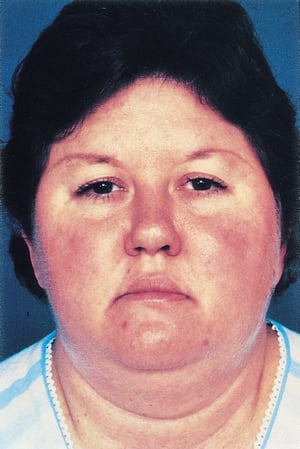
Moon face with a plethoric appearance
Truncal obesity with prominent supraclavicular and dorsal cervical fat pads (buffalo hump)
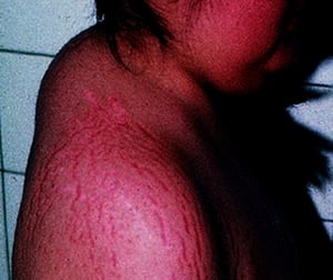
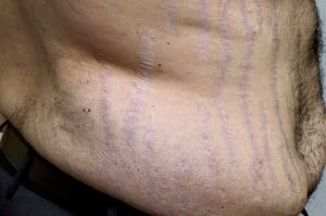
Striae (stretch marks)
Usually, very slender distal extremities and fingers
© Springer Science+Business Media
© Springer Science+Business Media
SCIENCE PHOTO LIBRARY

By permission of the publisher. From Biller B. In Atlas of Clinical Endocrinology: Neuroendocrinology and Pituitary Disease. Edited by SG Korenman (series editor) and ME Molitch. Philadelphia, Current Medicine, 2000.


© Springer Science+Business Media


© Springer Science+Business Media


SCIENCE PHOTO LIBRARY


By permission of the publisher. From Biller B. In Atlas of Clinical Endocrinology: Neuroendocrinology and Pituitary Disease. Edited by SG Korenman (series editor) and ME Molitch. Philadelphia, Current Medicine, 2000.
Muscle wasting and weakness are present. The skin is thin and atrophic, with poor wound healing and easy bruising. Striae may appear on the abdomen. Hypertension, renal calculi, osteoporosis, glucose intolerance, reduced resistance to infection, and mental disturbances are common. Cessation of linear growth is characteristic in children.
Females usually have menstrual irregularities. In females with adrenal tumors, increased production of androgens may lead to hirsutism, temporal balding, and other signs of virilism.
Diagnosis of Cushing Syndrome
Urinary free cortisol level
Dexamethasone suppression test
Midnight serum or salivary cortisol levels
Plasma ACTH levels; if detectable, provocative testing
Diagnosis is usually suspected based on the characteristic symptoms and signs. Confirmation (and identification of the cause) generally requires hormonal and imaging tests.
Urinary free cortisol measurement
In some centers, testing begins with a 24-hour measurement of urinary free cortisol, which is elevated > 120 mcg/24 hours (> 331 nmol/24 hours) in almost all patients with Cushing syndrome. However, many patients with elevations of urinary free cortisol between 100 and 150 mcg/24 hours (276 and 414 nmol/24 hours) have obesity, depression, or polycystic ovaries but not Cushing syndrome. Normal ranges may vary according to assay.
A patient with suspected Cushing syndrome with grossly elevated urinary free cortisol (> 4 times the upper limit of normal) almost certainly has Cushing syndrome. Two to 3 normal collections usually exclude the diagnosis. Slightly elevated levels generally necessitate further investigation, as do normal levels when clinical suspicion is high.
A baseline morning (eg, 9 AM) serum cortisol measurement should also be done.
Dexamethasone suppression test
dexamethasone is given orally at 11 to 12 PM and serum cortisol is measured at 8 to 9 AM the next morning. In most normal patients, this medication suppresses morning serum cortisol to < 1.8 mcg/dL (< 50 nmol/L), whereas patients with Cushing syndrome virtually always have a higher level. A more specific but equally sensitive test is to give dexamethasone 0.5 mg orally every 6 hours for 2 days (low dose). In general, a clear failure to suppress cortisol levels in response to low-dose dexamethasone establishes the diagnosis, unless there is reason to suspect abnormal dexamethasone absorption or metabolism.
Midnight cortisol measurements
If results of urinary free cortisol measurements and the dexamethasone suppression test are indeterminate, the patient can be hospitalized for measurement of serum cortisol at midnight, which is more likely to be conclusive. Alternatively, and more conveniently, the patient may collect salivary cortisol samples and store them in the refrigerator at home. Serum cortisol normally ranges from 5 to 25 mcg/dL (138 to 690 nmol/L) in the early morning (6 to 8 AM) and declines gradually to < 1.8 mcg/dL (< 50 nmol/L) at midnight. Patients with Cushing syndrome occasionally have a normal morning serum cortisol level but lack normal diurnal decline in cortisol production, such that the midnight serum cortisol levels are above normal and the total 24-hour cortisol production may be elevated. Normal ranges of midnight salivary cortisol level vary according to assay.
Serum cortisol may be spuriously elevated in patients with congenital increases of corticosteroid-binding globulin or in those receiving estrogen therapy, but diurnal variation is normal in these patients.
Plasma ACTH measurement
ACTH levels are measured to determine the cause of Cushing syndrome. Undetectable levels suggest a primary adrenal cause. High levels suggest a pituitary cause or an ectopic source. If ACTH is detectable, provocative tests help differentiate Cushing disease from ectopic ACTH syndrome, which is rarer.
In response to high-dose dexamethasone (2 mg orally every 6 hours for 48 hours), the 9 AM serum cortisol falls by >Diagnostic Tests in Cushing Syndrome). Corticotropin-releasing hormone (CRH) causes a similar rise in ACTH and cortisol and has been used in diagnostic testing; however, its availability is limited.
> 3 virtually excludes ectopic ACTH syndrome, whereas a ratio < 3 suggests a need to seek such a source.
Imaging
In children with Cushing disease, pituitary tumors are very small and usually cannot be detected with MRI. Petrosal sinus sampling is particularly useful in this situation. MRI is preferred to CT in pregnant women to avoid fetal exposure to radiation.
Treatment of Cushing Syndrome
Surgery or radiation therapy to remove pituitary, adrenal, or ectopic ACTH-producing tumors
Sometimes somatostatin analogs or dopamine
Ketoconazole
ACTH-secreting pituitary tumors
Pituitary tumors that produce excessive ACTH are removed surgically or extirpated with radiation therapy. If no tumor is shown on imaging but a pituitary source is likely, total hypophysectomy may be attempted, particularly in older patients. Younger patients (including children and adolescents) may receive supervoltage irradiation of the pituitary, delivering 45 Gy (Gray). However, in children, irradiation may reduce secretion of growth hormone and occasionally cause precocious puberty. In special centers, a focused beam of radiation therapy may be given as a single dose (radiosurgery). Alternatively, proton beam therapy can be used if available. Response to irradiation occasionally requires several years, but response is more rapid in children.
dopaminepasireotidemifepristone increases serum cortisol but blocks the effects of the corticosteroid and may cause hypokalemia.
Bilateral adrenalectomy is reserved for patients with pituitary hyperadrenocorticism who do not respond to both pituitary exploration (with possible adenomectomy) and irradiation, or in patients in whom surgery was unsuccessful and radiotherapy is contraindicated. Adrenalectomy requires life-long corticosteroid replacement.
Corticosteroid-secreting adrenocortical tumors
Adrenocortical tumors are removed surgically. Patients must receive cortisol during the surgical and postoperative periods because their nontumorous adrenal cortex will be atrophic and suppressed.
Benign adenomas can be removed laparoscopically.
With multinodular adrenal hyperplasia, bilateral adrenalectomy may be necessary, but in some cases removal of the larger adrenal alone may be effective. Even after a presumed total adrenalectomy, functional regrowth occurs in a few patients.
Ectopic ACTH-producing tumors
In the case of a disseminated ACTH-producing carcinoid or other neuroendocrine tumor, bilateral adrenalectomy nay be indicated.
> 2 years requires close follow-up because mild gastritis, gallstones, cholangitis, and malabsorption may develop.
Nelson syndrome
Nelson syndrome occurs when the pituitary gland continues to expand after bilateral adrenalectomy, causing a marked increase in the secretion of ACTH and its precursors and resulting in severe hyperpigmentation. It occurs in approximately 20 to 25% of patients who undergo adrenalectomy (1). The risk is probably reduced if the patient undergoes prophylactic pituitary radiation therapy at the time of adrenalectomy, but most centers would simply scan the pituitary at frequent intervals with regular assessment of ACTH levels.
Although irradiation may arrest continued pituitary growth, many patients also require hypophysectomy. The indications for hypophysectomy are the same as for any pituitary tumor: an increase in size such that the tumor encroaches on surrounding structures, causing visual field defects, pressure on the hypothalamus, or other complications.
Radiation therapy may be given if it was not given at the time of bilateral adrenalectomy. Radiosurgery, or focused radiation therapy, can be given in a single fraction when standard external beam radiation therapy has already been done, as long as the lesion is at a reasonable distance from the optic nerve and chiasm.
Treatment reference
1. Reincke M, Albani A, Assie G, et al. Corticotroph tumor progression after bilateral adrenalectomy (Nelson's syndrome): systematic review and expert consensus recommendations. Eur J Endocrinol 2021;184(3):P1-P16. doi:10.1530/EJE-20-1088
Key Points
Diagnosis is usually made by elevated nocturnal serum or salivary cortisol levels, or 24-hour urinary free cortisol level, and a dexamethasone suppression test in which serum cortisol does not suppress.
Pituitary causes are distinguished from nonpituitary causes by adrenocorticotropic hormone (ACTH) levels.
Imaging is then done to identify any causative tumor.
Tumors are usually treated surgically or with radiation therapy.


